Syndesmosis sprains : The high ankle injury
You may have heard various athletes suffering a high ankle sprain or injuring their syndesmosis. But what exactly is a syndesmosis injury? And how does it differ to a normal lateral ankle sprain?
The ankle syndesmosis is the joint between the distal (lowest aspect) of your tibia and fibula. It is comprised by three main supporting ligamentous structures – The Anterior inferior tibiofibular ligament, Posterior inferior Tibiofibular ligament, and interosseous membrane (see Figure 1). The role of the syndesmosis is to provide stability to the tibia and fibula and resist separation of these two bones during weightbearing tasks. It also plays a role in assisting with mobility of the ankle.
You may have heard various athletes suffering a high ankle sprain or injuring their syndesmosis. But what exactly is a syndesmosis injury? And how does it differ to a normal lateral ankle sprain?
The ankle syndesmosis is the joint between the distal (lowest aspect) of your tibia and fibula. It is comprised by three main supporting ligamentous structures – The anterior inferior tibiofibular ligament, posterior inferior tibiofibular ligament, and interosseous membrane (see Figure 1). The role of the syndesmosis is to provide stability to the tibia and fibula and resist separation of these two bones during weightbearing tasks. It also plays a role in assisting with mobility of the ankle.
How does it differ to a common ankle sprain?
Generally, a lateral ankle sprain is a result of and inversion injury and will result in an injury to the outside ligaments of your ankle (ATFL, CFL, PTFL). These ligaments are positioned slightly lower than the syndesmosis and provide stability to the true ankle joint.
Mechanisms of injury:
The most common mechanism for injuring your syndesmosis is a forced dorsiflexion combined with an Eversion movement. Essentially the foot/ankle moves in an upward direction and to the outside of the leg (See figure 3).
The syndesmosis can also be injured with a typical inversion or lateral ankle sprain (Figure 2) mechanism. This usually occurs when the incident is of high force and will result with an injury to the lateral ligaments as well.
Signs and symptoms:
· Mechanism of injury consistent with a syndesmosis injury (forced dorsiflexion + Eversion)
· Pain location may extend above the ankle and into the lower shin
· Swelling may sit slightly above the cease line of the ankle joint
· Difficulty weightbearing, particularly when the foot is in dorsiflexion (knee over toe)
· Low confidence/feeling of instability
Gradings:
Grade 1: isolated injury to the AITFL
Grade 2: Injury to the AITFL and interosseous membrane
Grade 3: Injury to the AITFL, interosseous membrane and PITFL
Grade 4: Injury to the AITFL, interosseous membrane, PITFL and deltoid ligament
Immediate management:
As always if you have recently suffered an injury, please seek medical attention from your physio or doctor for accurate diagnosis and management.
If a syndesmosis injury is suspected acute management will initially involve offloading and protecting the tissues. This may be in the form of one or a combination of crutches, a cam walker (moon) boot and strapping.
Your physio or Doctor may also refer you for imaging such as an x-ray or MRI to assist with diagnosis and understanding the severity of the injury.
Following the acute period of offloading and protection a period of rehabilitation will be required to restore normal function of the foot and ankle. In more severe cases surgery may be required to stabilise the syndesmosis and therefore rehab will commence following a period of protection post-surgery.
If you have experienced an ankle sprain yourself, please book in with one of our physiotherapists for an individualised rehabiltation program.
Early Stage Ankle Sprain Rehabilitation
Ankle sprains are one of the most common lower limb injuries reported by active individuals, with a high reoccurrence rate. The lateral ligaments (outside of the ankle) are the most commonly injured, as discussed in one of our previous blogs as seen here https://www.eastvicparkphysiotherapy.com.au/news/2021/1/14/chronic-ankle-instability
Injury prevention and rehabilitation is an effective way to reduce the risk of post injury recurrence.
Key areas of a rehab plan include the following
Restoring full range of movement
Restoring range of motion is important in the initial stages of rehab, this can be achieved by correct heel toe walking (if needed with the assistance of crutches dependant on severity of injury). These exercises are used in the beginning phase of rehabilitation
Ankle Active range of motion
- Ankle Alphabets
- Ankle Pumps
- Calf Stretching
Pain free stationary cycling is also a great way to progress active range of motion exercises as well as re introducing a cardiovascular component to the program.
Muscle Strength
Strength needs to be addressed in all directions available in the ankle. These include dorsiflexion, plantar flexion, inversion, eversion. To increase the difficulty of these movements, your physiotherapist may use external resistance, such as therabands, or using your own body weight, through calf raise exercise. Body weight exercises are encouraged as soon as the injury is pain free.
Proprioception
Proprioception is the awareness of joint position and movement, and this becomes impaired after a ligament injury. It is an important part of ankle injury rehabilitation and can start early in your program. Examples of proprioception exercises include:
- Standing on one leg
- Balance Boards
The above exercises are only a guide and will need to be progressed to ensure a full recovery. If you have experienced an ankle sprain please book in with one of our physiotherapists to have your rehabilitation individualised to suit your needs.
THE IMPORTANCE OF MUSCULOSKELETAL SCREENING
Finals time for most winter sports is fast approaching and from a physiotherapy perspective this is the time of year that we see a spike in sporting injuries. A lot of these injuries tend to be to parts of the body that have some sort of deficit, be it strength, length or control. It is quite hard to be able to identify these areas yourself and even physiotherapists would find it hard to accurate identify these deficits purely through observation.
Finals time for most winter sports is fast approaching and from a physiotherapy perspective this is the time of year that we see a spike in sporting injuries. A lot of these injuries tend to be to parts of the body that have some sort of deficit, be it strength, length or control. It is quite hard to be able to identify these areas yourself and even physiotherapists would find it hard to accurate identify these deficits purely through observation.
This is why screening is so widely utilised for athletes from amateur to elite. Screening usually involves a battery of tests that give objective measurements that are then compared to the normal values for an athlete in a specific sport. Screening can also involve questionnaires that focus on general health and previous injury history.
An article by Sanders, Blackburn and Boucher (2013), looked at the use of pre-participation physicals (PPE) for athletic participation. They found PPE’s to be useful, comprehensive and cost effective. They explained that PPE’s can be modified to meet the major objectives of identification of athletes at risk.
An article by Maffey and Emery (2006) looked at the ability of pre-participation examinations to contribute to identifying risk factors for injury. They found limited evidence for examinations in terms of the ability to reduce injury rates among athletes. However, they were effective in the identification of previous injury (such as ankle sprains) and providing appropriate prevention strategies (such as balance training). From this it has been shown to reduce the risk of recurrent injury. It may also be useful in identifying known risk factors which can be addressed by specific injury prevention interventions.
An example of a screening measure that is typically used in screening protocols includes a knee to wall test (KTW). This test is used for ankle dorsiflexion as well as soleus muscle length (one of your calf muscles). The test is performed using a ruler which is placed perpendicular to a wall with no skirting board. The athlete puts their foot flat on the ground next to the ruler and as far from the wall as possible as long as their knee is touching the wall. Distance from the wall to the end of the big toe is noted by looking at the ruler. An example of a normal distance for netball players is greater than 15cm on each side.
Here at East Vic Park Physiotherapy we have developed a number of specific musculoskeletal screens for a variety of sports including netball, running, swimming and throwing sports. They comprehensively identify the key risk factors that are seen in injuries sustained in each sport. If you are interested in preventing injury for the upcoming sports season, then contact the clinic on 9361 3777 and book your screening appointment today!
Plantar fasciitis
WHAT IS IT?
Plantar fasciitis Is a very common cause of heel pain. It can be quite debilitating and can last for months if not addressed. Typically, pain will be felt on the inside of the heel and arch. Pain can be sharp or achy. There can be a small amount of swelling over the medial heel as well as tenderness to touch. Mornings are worse, with it usually taking anywhere from 2-3 minutes to an hour for the stiffness and pain to reduce.
POSSIBLE CAUSES
· Change in load eg Running/jumping
· Change in footwear
· Change in activity surface eg. Hard surface
· Acute trauma eg. Stepping on a rock
SCANS
Sometimes your GP will refer you for a scan of the affected area. Most likely it will be an x-ray or an ultrasound. This may show that there are heel spurs or “tears” in the plantar fascia. Although it can be good to confirm the diagnosis, scans can sometimes be detrimental as it may cause people to become worried about their condition. Scan results can also correlate poorly with symptoms an example being that people with heel spurs on x-ray don’t necessarily develop Plantar fasciitis.
TREATMENT OPTIONS
· Soft tissue release
· Joint mobilisations
· Taping techniques
· Orthotics
· Exercise program (Physiotherapist prescribed)
· Load management plan (Physiotherapist prescribed)
LOAD MANAGEMENT
Load management is about controlling how much you use the particularly area on a day to day basis. Usually when an area becomes painful, its load capacity (ability to tolerate load) is reduced so it becomes overloaded quicker than normal. This means that even normal tasks or activities like walking or standing can cause it to become more painful and swollen.
One of the ways to improve the capacity is to progressively build up the amount that you use that area. This can be done with a specific structured exercise program (physiotherapist prescribed) that is made more difficult over a period of time. It is normal for rehabilitation to be painful, you cannot improve load tolerance without causing some discomfort.
The best way to monitor improvement is by recording morning pain (rating it out of 10, 10 is worst, 0 is nothing). It is normal to have ongoing morning stiffness even after pain has completely disappeared.
DIFFERENTIAL DIAGNOSIS
Sometimes Plantar fasciitis might not be the cause of heel or foot pain. It is important to see a physiotherapist to get an accurate diagnosis. Other causes of heel pain are below:
· Plantar or Calcaneal Nerve pain
· S1 radiculopathy
· Stress fracture
· Tarsal tunnel syndrome
· Fractures
· Retrocalcaneal bursitis
· Spondyloarthropathies
· Cancer (osteoid osteoma)
TIPS FOR PAIN FLARE UPS
· Try to avoid walking around in bare feet
· Using ice over the sore area can give temporary relief
· Stretching it may be uncomfortable so roll a golf ball/tennis ball under the foot instead to release tight muscles
· Pain relief or anti-inflammatory medication can be helpful but ask your pharmacist for advice
· See your physiotherapist for a progressive loading program
UNDERSTANDING PAIN
An excellent video on what our understanding of pain currently is and in particular the complexities of chronic pain.
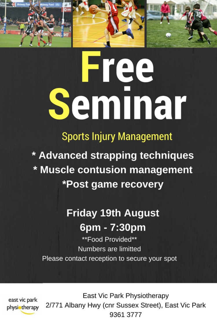
SPORTS INJURY MANAGEMENT SEMINAR
Whether your sports season is heading into finals or you are about to start gearing up for the summer season ahead, the information presented will help you to perform at your best.
a FREE seminar on sports injury management presented by the Physiotherapists at East Vic Park Physiotherapy. Topics will include muscle contusion (corkie) management, post-game recovery and a practical session on strapping.
Whether your sports season is heading into finals or you are about to start gearing up for the summer season ahead, the information presented will help you to perform at your best.
Appropriate for all athletes, parents, trainers and coaches.
Food will be provided - let us know if you have any dietary requests.
Spaces are limited so call us on 9361 3777 to secure your place now.


