How to classify muscle injuries on MRI using the British Athletic Muscle Injury Classification (BAMIC) system
Soft tissue injuries are very common in sport/physical activity and recovery timelines can take anywhere from 10 days – 16 weeks. These timelines are decided using physical assessment and understanding of muscle-tendon complex injury healing but, in the case of lower limb injuries, can be aided with an MRI of the area. The radiologists who interpret the scan results will often use the BAMIC system to identify the area and grade of the injury which can help in determining return to play timelines.
Soft tissue injuries are very common in sport/physical activity and recovery timelines can take anywhere from 10 days – 16 weeks. These timelines are decided using physical assessment and understanding of muscle-tendon complex injury healing but, in the case of lower limb injuries, can be aided with an MRI of the area. The radiologists who interpret the scan results will often use the BAMIC system to identify the area and grade of the injury which can help in determining return to play timelines.
The area is typically divided into three parts of the muscle-tendon complex:
1. Myofascial: Involves the periphery of the muscle, including the surrounding connective tissue layers (epimysium, perimysium, aponeurosis). The myofascial allows force transmission and absorption through the muscle-tendon unit.
2. Myotendinous junction/muscular: A blend of muscle and tendon connection this is potentially the location of greatest force production within the muscle tendon unit.
3. Tendon: The most serious of the three when the central tendon is involved as there are high re-injury rates if the person returns to high level physical activity too early. Tendons take longer to heal than muscle or fascia due to the less vascularity. Also important to note that intra-muscular tendon parts has less sensory neural innervation which means reported pain levels is not a good indicator of severity of injury with those athletes.
Classification system
grade 0a: focal neuromuscular injury with normal MRI
grade 0b: generalised muscle soreness with normal MRI or MRI findings typical of delayed onset muscle soreness (DOMS)
grade 1 (mild): high STIR signal that is <10% cross-section or longitudinal length <5 cm with <1 cm fibre disruption
grade 2 (moderate): high STIR signal that is 10-50% cross-section; longitudinal length 5-15 cm with <5 cm fibre disruption
grade 3 (extensive): high STIR signal that is >50% cross-section or longitudinal length >15 cm with >5 cm fibre disruption
grade 4: complete tear
Rehabilitation and return to play considerations
The health professional will typically take into account a myriad of factors to get someone back to exercise as quickly (and safely) as possible. These include (but are not limited to):
· Previous injury history
· Physical assessment findings
· Current activity level
· Goals of the patient
· Response to treatment and rehab
· Time of the season (eg finals etc)
Here at East Vic Park, we are experienced in treating soft tissue injuries. Book in to see one of your friendly physiotherapists today.
Syndesmosis sprains : The high ankle injury
You may have heard various athletes suffering a high ankle sprain or injuring their syndesmosis. But what exactly is a syndesmosis injury? And how does it differ to a normal lateral ankle sprain?
The ankle syndesmosis is the joint between the distal (lowest aspect) of your tibia and fibula. It is comprised by three main supporting ligamentous structures – The Anterior inferior tibiofibular ligament, Posterior inferior Tibiofibular ligament, and interosseous membrane (see Figure 1). The role of the syndesmosis is to provide stability to the tibia and fibula and resist separation of these two bones during weightbearing tasks. It also plays a role in assisting with mobility of the ankle.
You may have heard various athletes suffering a high ankle sprain or injuring their syndesmosis. But what exactly is a syndesmosis injury? And how does it differ to a normal lateral ankle sprain?
The ankle syndesmosis is the joint between the distal (lowest aspect) of your tibia and fibula. It is comprised by three main supporting ligamentous structures – The anterior inferior tibiofibular ligament, posterior inferior tibiofibular ligament, and interosseous membrane (see Figure 1). The role of the syndesmosis is to provide stability to the tibia and fibula and resist separation of these two bones during weightbearing tasks. It also plays a role in assisting with mobility of the ankle.
How does it differ to a common ankle sprain?
Generally, a lateral ankle sprain is a result of and inversion injury and will result in an injury to the outside ligaments of your ankle (ATFL, CFL, PTFL). These ligaments are positioned slightly lower than the syndesmosis and provide stability to the true ankle joint.
Mechanisms of injury:
The most common mechanism for injuring your syndesmosis is a forced dorsiflexion combined with an Eversion movement. Essentially the foot/ankle moves in an upward direction and to the outside of the leg (See figure 3).
The syndesmosis can also be injured with a typical inversion or lateral ankle sprain (Figure 2) mechanism. This usually occurs when the incident is of high force and will result with an injury to the lateral ligaments as well.
Signs and symptoms:
· Mechanism of injury consistent with a syndesmosis injury (forced dorsiflexion + Eversion)
· Pain location may extend above the ankle and into the lower shin
· Swelling may sit slightly above the cease line of the ankle joint
· Difficulty weightbearing, particularly when the foot is in dorsiflexion (knee over toe)
· Low confidence/feeling of instability
Gradings:
Grade 1: isolated injury to the AITFL
Grade 2: Injury to the AITFL and interosseous membrane
Grade 3: Injury to the AITFL, interosseous membrane and PITFL
Grade 4: Injury to the AITFL, interosseous membrane, PITFL and deltoid ligament
Immediate management:
As always if you have recently suffered an injury, please seek medical attention from your physio or doctor for accurate diagnosis and management.
If a syndesmosis injury is suspected acute management will initially involve offloading and protecting the tissues. This may be in the form of one or a combination of crutches, a cam walker (moon) boot and strapping.
Your physio or Doctor may also refer you for imaging such as an x-ray or MRI to assist with diagnosis and understanding the severity of the injury.
Following the acute period of offloading and protection a period of rehabilitation will be required to restore normal function of the foot and ankle. In more severe cases surgery may be required to stabilise the syndesmosis and therefore rehab will commence following a period of protection post-surgery.
If you have experienced an ankle sprain yourself, please book in with one of our physiotherapists for an individualised rehabiltation program.
FIVE questions you should ALWAYS ask your orthopaedic surgeon before making a decision on managemen
Orthopedic surgeons are indispensable members of the health profession and have a level of anatomical and biomechanical knowledge and a degree of experience that is rarely surpassed.
However, if you’ve ever been for a consultation with an orthopedic surgeon, you’ll know how fast the appointment passes. Many of our patients have reported being in-and-out without feeling like they’ve said more than 10 words.
Most consults will last 10-15 minutes and within this short space the surgeon has a certain “volume” of assessment to conduct and information they MUST deliver. It’s easy to see why they can occupy most of consultation to you leaving little opportunity for questions or express your opinion. When the opportunity finally arises, many patients are so bombarded with information that they forget the questions that had been circling in their head for days!
The surgeon is not to blame here as they are exceptionally busy individuals and with a huge demand on their limited time and wealth of knowledge and experience.
However, it makes it imperative that you use the time well and ask direct and concise questions to ensure you leave the session fully informed and able to make the decision that is best for you in your unique situation.
We decided to put together a list of questions that you should leave the consultation with the answers to. We’d recommend you print the 5 questions (and add any others you think of) and review them quickly before leaving the consultation to ensure you don’t leave with unaddressed concerns.
1. Is there any way that I can AVOID surgery and how would outcomes compare if I took this option?
ALWAYS ASK THIS QUESTION.
There is ALWAYS an alternative when it comes to surgery.
For example, considering a shoulder dislocation in a young male AFL player, a stabilisation surgery is highly recommended. Without surgery, this patient would have at least a 70% risk of the injury recurring. In contrast, with a Latarjet stabilisation he could expect as low as a 2.7% risk of recurrence.
However, you always have choices. You could try and be one of the 30% who survive non-operatively. Likewise some patients might have no desire to return to AFL and consider moving to a less “risky” sport such as triathlons where an operation is completely unnecessary. So even though we would recommend the stabilisation surgery in general, for 3 different people, they may choose 3 different options based on their own preferences.
To sum up, there is never a single option and you need to ask this question to have accurate information to allow you to weigh up the positives and negatives of all your options and make the right choice for you.
2. How long CAN it take to recover - worst case scenario? What will I feel/how painful will it be?
Yes, the problem/pathology may be fixed with surgery, but the soft tissue that is disrupted in the process will be painful and take some time to recover from. This obviously depends on the nature of the surgery but it will almost always be very painful initially.
Everyone asks how long it will take to recover. The surgeon will often tell you how long it usually takes based on average outcomes.
However, humans are complex and varied creatures, and so surgery is not like changing a part on a car.
For example, if a surgeon advises that you CAN start walking crutch-free at 7 days-post does not mean that you WILL. Some may be ready at 4 days, while some may take 14.
Many patients get disheartened because they’re running behind the timeline, or because a friend had the same procedure and was much better at this point. However, the timeline is based on the average recovery, and your friend may be an unusually high performer due to a whole host of factors such as severity of condition, genetics and specific surgical differences.
To manage these complex situations, it’s always a good option to ask the surgeon how long it CAN take and ask for the worst case scenario. It’s important to not catastrophize about this as it is an unlikely outcome, but knowing it can help prevent frustration when your’re running behind the average timeline. This also gives us a clear definition to differentiate between things are going slowly and things are going wrong.
3. Are there other ways to do this surgery? Why have we chosen this variation?
This question will get a bit technical and some may prefer not to worry about it, but there are many ways of surgically achieving your goal.
Clinical trials help provide us with some answers as to which way is the most likely to be effective, but also provide an insight as to what can go wrong.
For example, to revisit shoulder dislocations, there are two surgical approaches most commonly used: the Latarjet procedure or an arthroscopic bankart repart.
There is now a solid amount of research indicating a higher rate of return to sport and a lower rate of recurrence of dislocation with a Latarjet. However, it is also associated with a slightly higher risk of adverse effects, so the decision is not always cut-and-dry.
Similarly, with and ACL reconstruction, choice of graft (hamstring, quad, patellar etc), tunnel location, single or double bundle, nerve block used can all effect outcome. Many factors influence the surgeons decision, and they will undoubtedly offer the best solution in their opinion. While you do no need to have personal knowledge on any of the above, we think it’s a good idea to obtain this information and understand WHY they have made the decisions they have.
It’s a good idea to write this down too as the technical terms are usually hard to recall.
4. How many of these procedures do you perform a year?
There is a body of research regarding total knee replacement operations and ACL repairs that consistently associates higher yearly volume (how many a year the surgeon performs) with fewer infections, shorter procedure time, shorter hospital stays, lower rate of transfusion, and better outcomes in the long-term. Research on other procedures is less available, but it’s safe to extrapolate that experience matters, just like any other profession.
If the surgeon has a relatively low yearly volume, it doesn’t mean that they will do a bad job as they will have undergone may procedures in their training and education.
Likewise, certain procedures as less commonly performed (e.g. repair of a complex bone break) so a high yearly volume is not feasible.
In any case, it’s worth asking how often they perform the procedure. If they perform a low number annually (<10 on a common procedure such as TKR), you could consider politely inquire if they have colleagues that specialize in this procedure and perform it more often.
With something as serious as a surgical procedure, you are always entitled to a second opinion. A good surgeon will have the self-awareness to know when they are and are not the right person for the job. If they can confidently assure you they have the necessary skill-set and experience, then you leave the consult knowing you are in safe hands.
5. Always ask YOURSELF, do I understand the diagnosis and/or the proposed solution? Test this by trying to summarize the information to you surgeon before you leave.
It’s a simple place to finish but make sure you fully understand what your surgeon has told you in the first place as errors in communication are so common in this situation.
The true test of this comes when you go home and try the “family/friend test”. If you can explain your problem well to family/friends, then you probably took the information in effectively. However, if it makes perfect sense in your head at the time, but you struggle to explain it later then you may not have understood it as well as you thought.
To ensure you pass the” family/friends test’, I recommend people try and perform a summary at the end of their consult, e.g.:
“So, if I understand correctly, my problem is that……and the proposed solution would fix this by…….all going well I should expect……but it may take as long as……..”.
This way, your surgeon will be able to highlight and correct anything that got lost in translation on the first occasion.
Conclusion:
There you have it – five important questions you should always ask in a consultation with an orthopedic surgeon. Any situation that requires the consideration of surgery is bound to be complex so it’s imperative that you get all the information. These five questions will help keep you focused and ensure you get the information needed to make a fully-informed decision that is right for you and your set of circumstances
THE IMPORTANCE OF MUSCULOSKELETAL SCREENING
Finals time for most winter sports is fast approaching and from a physiotherapy perspective this is the time of year that we see a spike in sporting injuries. A lot of these injuries tend to be to parts of the body that have some sort of deficit, be it strength, length or control. It is quite hard to be able to identify these areas yourself and even physiotherapists would find it hard to accurate identify these deficits purely through observation.
Finals time for most winter sports is fast approaching and from a physiotherapy perspective this is the time of year that we see a spike in sporting injuries. A lot of these injuries tend to be to parts of the body that have some sort of deficit, be it strength, length or control. It is quite hard to be able to identify these areas yourself and even physiotherapists would find it hard to accurate identify these deficits purely through observation.
This is why screening is so widely utilised for athletes from amateur to elite. Screening usually involves a battery of tests that give objective measurements that are then compared to the normal values for an athlete in a specific sport. Screening can also involve questionnaires that focus on general health and previous injury history.
An article by Sanders, Blackburn and Boucher (2013), looked at the use of pre-participation physicals (PPE) for athletic participation. They found PPE’s to be useful, comprehensive and cost effective. They explained that PPE’s can be modified to meet the major objectives of identification of athletes at risk.
An article by Maffey and Emery (2006) looked at the ability of pre-participation examinations to contribute to identifying risk factors for injury. They found limited evidence for examinations in terms of the ability to reduce injury rates among athletes. However, they were effective in the identification of previous injury (such as ankle sprains) and providing appropriate prevention strategies (such as balance training). From this it has been shown to reduce the risk of recurrent injury. It may also be useful in identifying known risk factors which can be addressed by specific injury prevention interventions.
An example of a screening measure that is typically used in screening protocols includes a knee to wall test (KTW). This test is used for ankle dorsiflexion as well as soleus muscle length (one of your calf muscles). The test is performed using a ruler which is placed perpendicular to a wall with no skirting board. The athlete puts their foot flat on the ground next to the ruler and as far from the wall as possible as long as their knee is touching the wall. Distance from the wall to the end of the big toe is noted by looking at the ruler. An example of a normal distance for netball players is greater than 15cm on each side.
Here at East Vic Park Physiotherapy we have developed a number of specific musculoskeletal screens for a variety of sports including netball, running, swimming and throwing sports. They comprehensively identify the key risk factors that are seen in injuries sustained in each sport. If you are interested in preventing injury for the upcoming sports season, then contact the clinic on 9361 3777 and book your screening appointment today!
Wrist and Hand Injuries
We use our hands repeatedly every day so it’s not surprising that sometimes we develop pain and discomfort in our fingers, wrists and forearms. Injuries in the wrist and hand can be caused due to traumatic events (e.g. a fall on an outstretched hand) or overuse, repetitive activities (e.g. computer use, racquet sports).
Anatomy
We use our hands repeatedly every day so it’s not surprising that sometimes we develop pain and discomfort in our fingers, wrists and forearms. Injuries in the wrist and hand can be caused due to traumatic events (e.g. a fall on an outstretched hand) or overuse, repetitive activities (e.g. computer use, racquet sports).
ANATOMY
The wrist and hand complex is made up of 27 bones, muscles, tendons, ligaments, nerves and blood vessels. Damage to any of these structures can cause pain and can affect your ability to use your hands effectively.
COMMON WRIST AND HAND INJURIES INCLUDE:
· Fractures
· Tendon pathologies e.g. Mallet finger, De Quervain’s Disease
· Ligament injuries e.g. sprains
· Joint inflammation
· Nerve entrapments e.g. Carpal tunnel syndrome
· Arthritic conditions
· Ganglion cysts
If hand and wrist injuries are not assessed and treated properly this may lead to further impairments in the future.
EARLY MANAGEMENT
Should include rest, ice, compression and elevation (RICE principle) for the first 48-72 hours. Anti-inflammatories (NSAIDS) also have a role in early management, taken in the form of tablets and topical gels.
PHYSIOTHERAPY
Your physiotherapist will go through a comprehensive assessment to determine the source of your pain. Once the source of your pain has been established the initial aim of treatment includes education and addressing acute symptoms (pain, lack of movement and loss of strength).
When indicated your physiotherapist will start to address other issues such as loading, muscle imbalances, poor posture and biomechanics.
PREVENTION
To reduce the risk of recurring wrist and hand injuries it is important to maintain adequate strength and length of the muscles around the wrist joint.
Your physiotherapist will advise you in activities that should be avoided to decrease irritation.
RETURN TO SPORT
When returning to sport it is essential that you discuss this with your physiotherapist. Your abilities will be assessed through a series of tests to determine whether you are ready to return to pre-injury activities and/or sport.
UNDERSTANDING PAIN
An excellent video on what our understanding of pain currently is and in particular the complexities of chronic pain.
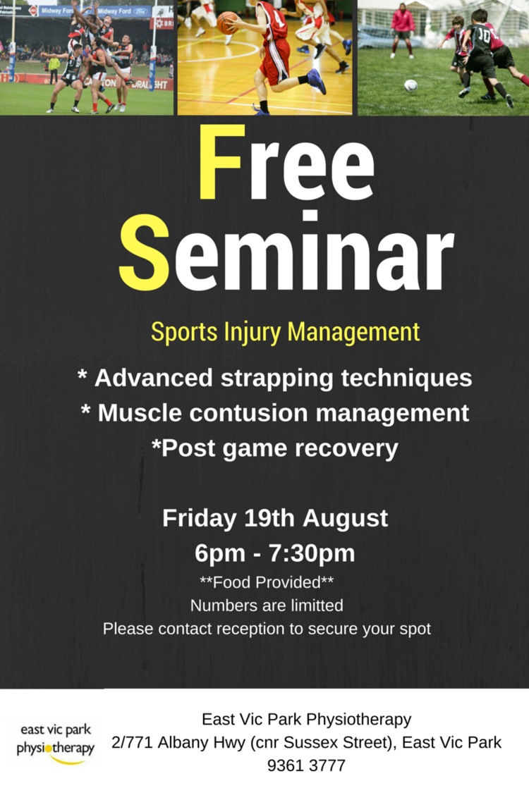
SPORTS INJURY MANAGEMENT SEMINAR
Whether your sports season is heading into finals or you are about to start gearing up for the summer season ahead, the information presented will help you to perform at your best.
a FREE seminar on sports injury management presented by the Physiotherapists at East Vic Park Physiotherapy. Topics will include muscle contusion (corkie) management, post-game recovery and a practical session on strapping.
Whether your sports season is heading into finals or you are about to start gearing up for the summer season ahead, the information presented will help you to perform at your best.
Appropriate for all athletes, parents, trainers and coaches.
Food will be provided - let us know if you have any dietary requests.
Spaces are limited so call us on 9361 3777 to secure your place now.


