How to classify muscle injuries on MRI using the British Athletic Muscle Injury Classification (BAMIC) system
Soft tissue injuries are very common in sport/physical activity and recovery timelines can take anywhere from 10 days – 16 weeks. These timelines are decided using physical assessment and understanding of muscle-tendon complex injury healing but, in the case of lower limb injuries, can be aided with an MRI of the area. The radiologists who interpret the scan results will often use the BAMIC system to identify the area and grade of the injury which can help in determining return to play timelines.
Soft tissue injuries are very common in sport/physical activity and recovery timelines can take anywhere from 10 days – 16 weeks. These timelines are decided using physical assessment and understanding of muscle-tendon complex injury healing but, in the case of lower limb injuries, can be aided with an MRI of the area. The radiologists who interpret the scan results will often use the BAMIC system to identify the area and grade of the injury which can help in determining return to play timelines.
The area is typically divided into three parts of the muscle-tendon complex:
1. Myofascial: Involves the periphery of the muscle, including the surrounding connective tissue layers (epimysium, perimysium, aponeurosis). The myofascial allows force transmission and absorption through the muscle-tendon unit.
2. Myotendinous junction/muscular: A blend of muscle and tendon connection this is potentially the location of greatest force production within the muscle tendon unit.
3. Tendon: The most serious of the three when the central tendon is involved as there are high re-injury rates if the person returns to high level physical activity too early. Tendons take longer to heal than muscle or fascia due to the less vascularity. Also important to note that intra-muscular tendon parts has less sensory neural innervation which means reported pain levels is not a good indicator of severity of injury with those athletes.
Classification system
grade 0a: focal neuromuscular injury with normal MRI
grade 0b: generalised muscle soreness with normal MRI or MRI findings typical of delayed onset muscle soreness (DOMS)
grade 1 (mild): high STIR signal that is <10% cross-section or longitudinal length <5 cm with <1 cm fibre disruption
grade 2 (moderate): high STIR signal that is 10-50% cross-section; longitudinal length 5-15 cm with <5 cm fibre disruption
grade 3 (extensive): high STIR signal that is >50% cross-section or longitudinal length >15 cm with >5 cm fibre disruption
grade 4: complete tear
Rehabilitation and return to play considerations
The health professional will typically take into account a myriad of factors to get someone back to exercise as quickly (and safely) as possible. These include (but are not limited to):
· Previous injury history
· Physical assessment findings
· Current activity level
· Goals of the patient
· Response to treatment and rehab
· Time of the season (eg finals etc)
Here at East Vic Park, we are experienced in treating soft tissue injuries. Book in to see one of your friendly physiotherapists today.
Most Common Injuries in Martial Arts
Martial Arts will leave you black and blue but we love it and here you’ll read about the most common injuries we see amongst fighters including some input from Australia’s most decorated fighter and all round top bloke John Wayne Parr.
Most Common Injuries in Martial Arts
Being a martial artist has its pro’s and con’s and injuries are a major risk of the job. The following are some common injuries that fighters can expect whether training for fun and fitness or aiming to become the world champ.
1. Lacerations
The most common injury in the MMA and martial arts world. Australia’s most decorated Muay Thai fighter John Wayne Parr currently counts a total of 346* stitches from 146 professional fights (Muay thai and boxing).
General rules for whether you need stitches are;
- If the cut is greater than 1-inch in length or depth
- If you can see fat, muscle or bone
- Is the cut easily exposed to infection i.e. on the hand or fingers
- Difficulty with clotting i.e. prolonged bleeding >15mins
- Is it in a sensitive area i.e. the hands or face where reducing scarring is integral.
See your GP or present to your nearest emergency department if you fit any of the above categories.
Good luck JWP in your next fight against Anthony ‘The Man’ Mundine.
2. Cork / Contusion / Haematoma
Is an impact on the muscle with sudden, heavy compressive force i.e. opposition players knee. These can present either within a muscle (intra-muscular) or between muscles (inter-muscular).
Inter-muscular corks are sore to touch, can bleed down the limb i.e. thigh towards the knee.
Intra-muscular cork are also tender, and stiff to touch where stretching is difficult and movements like walking can be difficult without a limp. These can sometimes lead to solidification of blood within the muscle if not resolved, known as ‘myositis ossificans’.
Management;
- 0-48hrs: involves rest, ice, compression and elevation post game.
- 2-3days: see a physio/doctor for assessment and whether anti-inflammatories will be of use. Start some gentle stretching and movement i.e. exercise bike or light training.
- 3-4 days: gentle massage around cork and training as tolerated.
- 5 days: stretching and foam rolling around the sore spot and gentle heat packs may be useful.
3. Concussion – Knock Out aka ‘the Big Kaboosh’ aka ‘goodnight Irene’
The risk of concussion is significant in MMA due to high volume of punches, elbows, knees and kicks to the head. However, many fights are stopped due to submissions as opposed to head injury. In February 2019, the Australian Institute of Sport, Australian Medical Association and Sports Medicine Australia released a joint Position Statement on the management of confirmed concussions in athletes. (https://ama.com.au/position-statement/concussion-in-sport-2019 ). This states that athletes over the age of 18 should not return to contact activity sooner than 7-days, and 18-year-olds and under should refrain from risk of contact for 14 days or more and once cleared by a doctor with no signs of concussion still present.
A player should be immediately removed from the sport if any of the following occurred;
- Loss of consciousness
- A fall to ground without protection
- Seizure or tonic (rigid) posturing
- Impaired memory (date, location)
- Severe headache and other symptoms (below)
- Vomiting
- Deteriorating consciousness
- Vision loss/changes
- Non-typical behaviour
Signs of concussion may include;
- Loss of consciousness, headache, drowsiness or feeling like ‘in a fog’ or ‘slow’
- Dizziness or balance problems
- Confusion, sadness, irritability, nervous, anxiousness or emotional
- Blurred vision or sensitivity to light or noise
- Trouble sleeping
If any of the symptoms above are severe then the person should be taken directly to emergency and/or immediately be assessed by a doctor.
4. Fractures
a. Boxers Fracture
i. Occurs when a closed fist impacts with another hard object (head/floor) causing fracture at the head of the metacarpal (typically the 5th metacarpal).
ii. Signs: Swelling, redness, tenderness or crunching (crepitus) over the affected area.
iii. Diagnosis: Doctors or physiotherapist physical exam and X-Ray
iv. Treatment: Non-surgical cast and immobility or surgical pins and immobilisation cast.
v. Prognosis: up to 12 weeks
Others – can include any part of the body due to the high forces of usually bone on bone impact.
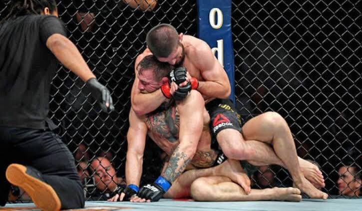
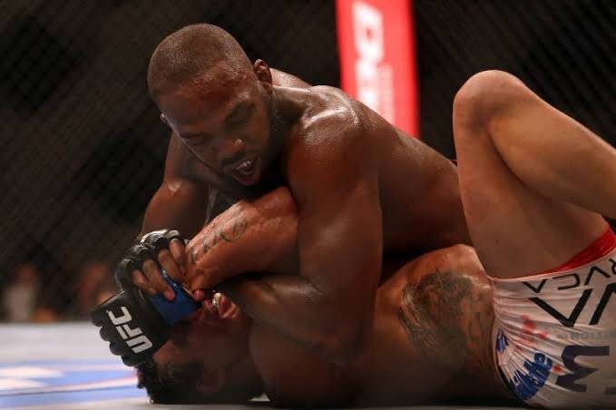
5. Cervical Disc Pathology – i.e. Neck cranks, rear-naked chokes.
Causes hyper flexion/extension or rotation to the neck. These often result in the fighter ‘tapping out’ prior to any serious damage. However multiple drilling, sparring and then fighting can place excessive stress on the neck discs, joints, ligaments and muscles.
Talk to your physiotherapist or GP immediately if you notice any of the following;
- Severe central neck pain
- Inability to move the neck >45° left or right
- Pins & needles, numbness or tingling into the arms and hands
- Severe shooting pains, electric shock or burning sensations in the arm and hands
- Strength loss in the upper limbs
Hot Tip: Learn your limits when training and ‘tap out’ early!
6. Dislocations / sub-luxations
The art of Brazilian Jiu Jitsu is designed to uncover the weakness at a joint and apply force so that the opponent taps before the trauma occurs. All dislocation/subluxations involve traumatic forces applied to a joint beyond its capability, leading to instability. Continuous micro-trauma may also lead to a joint becoming vulnerable over time.
A history of trauma puts you at increased risk of a repeat injury and continued clicking/locking/catching and instability indicates that the joint needs proper assessment and treatment. Never try to relocated a dislocated joint without a medical professional present.
Tip for young players… TAP EARLY
Author: Peter Gangemi - Master of Physiotherapy
Shoulder – Kimura / Americana
Shoulder – Kimura / Americana
Elbow – Arm bar
Thumb Dislocation
Compartment Syndrome: The muscle pain not to be missed!
Earlier this year, St Kilda football club’s caption, Jarryn Geary, sustained an innocuous quad corkie, which later developed into compartment syndrome requiring immediate emergency surgery. Compartment syndrome is a serious condition which can occur acutely or over time and often requires prompt medical attention. Do you know the signs and symptoms to look out for?
Compartment Syndrome is a condition where excessive pressure in a muscle compartment restricts blood supply to that muscle. Muscles are surrounded by fascial connective tissue, which has very poor elasticity. Excessive swelling in the fascial compartment results in compression of blood vessels, then resulting in a reduction of oxygen to that muscle.
Causes
Compartment Syndrome can occur acutely after a traumatic injury (eg fracture, contusion or surgery), or as the result of a blood clot. This is caused by ongoing swelling or bleeding entrapped in the muscle’s fascial casing, increasing pressure within the muscle compartment.
Occasionally compartment syndrome can have no apparent cause, and develops over weeks or months, triggered by vigorous exercise such as running or cycling. Symptoms of chronic compartment syndrome typically worsen throughout exercise and ease with rest.
Signs and Symptoms:
- Severe pain in the muscle
- Swelling or tightness
- Pale or waxy appearance of the skin (due to restricted blood supply)
- Pins and needles, burning or numbness
What do I do if I suspect compartment syndrome?
Get assessed as soon as possible by your physio or GP, particularly if you have a history of trauma to the area. Through careful assessment, your health professional will determine if compartment syndrome is likely, and will refer you on as necessary.
Acute compartment syndrome is considered a medical emergency and can be life threatening if not addressed promptly. Insufficient blood supply over a long period of time means the area is deprived of oxygen and can result in tissue and nerve necrosis (death).
Chronic compartment syndrome can be harder to diagnose, due to transient symptoms that ease with rest. A measurement of intracompartmental pressure can be used to determine the severity of swelling in the fascial compartment. A pressure higher than 30mmHg indicates excessive pressure in the compartment. A CT or MRI scan may be recommended to rule out other serious pathologies.
Treatment Options
Acute compartment syndrome is a medical emergency and a fasciotomy of the affected compartment must be performed immediately to prevent permanent tissue damage. This involves cutting the encasing fascia of the muscle to release pressure, decompress the blood vessels and restore blood flow to the area.
In chronic compartment syndrome, it is recommended trying conservative rehabilitation, firstly by reducing or ceasing physical exercise/activities that exacerbate symptoms. From there, a physiotherapist can assess your strength and biomechanics, to identify any factors that may influence the condition. A referral to a sports doctor may be appropriate to discuss medication options. If conservative rehabilitation fails, or if an individual would like to continue their sport at the same level of intensity, fasciotomy surgery needs to be considered.
Myositis Ossificans
WHAT IS IT?
Myositis ossificans (MO) is a condition where bone tissue forms within a muscle after injury. Damage of muscle tissue results in bleeding and inflammation within the first 24-48 hours post injury, and within the first week collagen fibres are laid down to repair the damaged tissue. Normally this process takes between 4-6 weeks to complete. With MO, fibrotic tissue is laid down in the muscle, which becomes ossified or calcified tissue.
This results in a hard, palpable lump in the muscle belly, restricted range of motion and reduced strength of the muscle. Unlike an uncomplicated muscle strain, MO can take months to years for the ossified portion to completely reabsorb. Return of muscle function is very dependable on the size of the ossification.
It is most common in the quadriceps, hamstring and back muscles.
HOW DOES IT HAPPEN?
The most common cause of myositis ossificans is significant, direct trauma to the muscle. It is most common in contact sports, however sports such as hockey and cricket also have a high prominence due to contact from the ball. It can also occur after a muscle strain.
You have increased risk of developing MO if you continue playing after a muscle injury, aggressively stretch or massage a muscle injury within the first 48 hours or re-injure the same area.
WHAT ELSE COULD IT BE?
A thorough clinical assessment will rule out any differential diagnosis however, particularly with a history of trauma, MO is hard to miss.
Medical imaging may be useful, however is not necessary to confirm a diagnosis. Ossified tissue can be seen on X-ray after 6 weeks, however if the injury is acute an ultrasound may be used to assess the extent of any haematoma.
Potential differential diagnosis that should be negated by your clinician are:
- Muscle strain
- Muscle contusion/haemotoma
- Joint sprain
- Compartment syndrome
HOW LONG WILL IT TAKE TO GET BETTER?
Over time, the ossification is gradually reabsorbed, however this process may take a number of months. In some cases the ossified growth may never completely reabsorb. This may result in tightness or discomfort when stretching the muscle, however normally won’t impair function of the muscle.
Here are some tips for the duration of your recovery. Your physiotherapist will guide you through an individual rehabilitation program.
Ice/compression
As with most acute injuries, the ‘RICE’ principle should be applied over the first 48 hours. This refers to rest, ice, compression and elevation. Compression can be applied with a tubigrip bandage, and icing for no longer than 20 minutes at a time, with a 2 hours break between ice packs.
Hirudoid cream
Hirudoid cream or gel dissolves small blood clots and reduces inflammation of the affected area. Depending on the area, the cream can be applied overnight and wrapped in gladwrap. It is advised to discuss with your pharmacist or GP before using this medication.
Load Management
Depending on the location and size of injury, you will need to de-load the area for a period of time. If you have significant discomfort walking, you may require crutches for the first few days, before you can tolerate putting weight on your leg. Your physio will then guide you through a program progressing through range of motion, strength, flexibility and power exercises. Unlike a muscle strain, it is okay (and often necessary) to push through slight discomfort with these exercises to regain full range and power of the muscle.
Stretching
An ossified section of the muscle tissue should be vigorously stretched so to not loose length of the affected muscle. Stretching should be performed for 2-3 minutes at a time, multiple times throughout the day. Note that stretching should not be performed within the first two weeks of a muscle injury as it can increase bleeding and should only be performed once the diagnosis of myositis ossificans is confirmed.
Returning to Play
Your physiotherapist should have a strict return to play criteria to ensure there is little risk of re-injury to the area. In contact sports you can play with padding over the area, to distribute the force of subsequent contact. It is oaky to return to play without complete reabsorption of the ossified area, as long are you have met the return to play criteria.
WHAT ELSE COULD I DO?
Anti-inflammatories may be indicated to help the body reabsorb ossified tissue. Your physiotherapist may refer you to a GP or sports physician to prescribe appropriate anti-inflammatories.
Surgical removal of the ossification is rare, and will only occur in those cases near a muscle insertion, where joint function is impaired. Surgery is only performed at least 12 months post original injury.
If you suspect myositis ossificans, your physiotherapist will help you develop a suitable management plan


